Shoulder Anatomy Diagram - Muscles Of The Pectoral Girdle And Upper Limbs Anatomy And Physiology / Illustration of the shoulder anatomy and labrum.
Dapatkan link
Facebook
X
Pinterest
Email
Aplikasi Lainnya
Shoulder Anatomy Diagram - Muscles Of The Pectoral Girdle And Upper Limbs Anatomy And Physiology / Illustration of the shoulder anatomy and labrum.. To further reinforce the shoulder, the four muscles of the rotator cuff extend from the scapula and surround the head of the humerus to both rotate the arm and prevent dislocation. Other important bones in the shoulder include: Plus, exercises for training them. These muscles are seen from the front (fig. With almost 360 degrees of motion (across several planes), the shoulder is by far the most mobile joint in the human body.
Rotator cuff injuries are very common, affecting over 3 million people in the united states every year. It has achieved this generous range of motion through the sacrifice of stability and a reliance on active and passive restraints (muscle, capsule and ligaments. The socket of the shoulder joint is shallow, and the labrum gives the socket more depth, and thus more stability. Diagram of a long bone anatomy. The shoulder is made up of three bones:
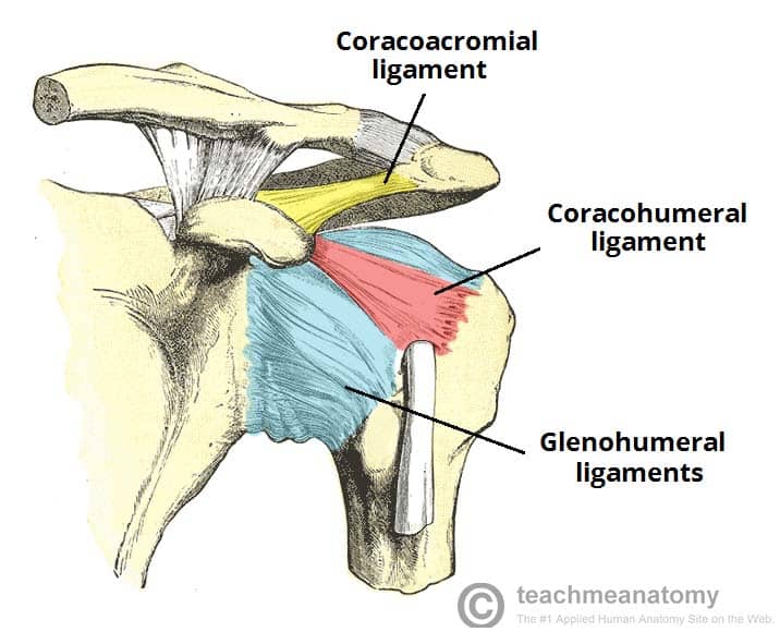
The Shoulder Joint Structure Movement Teachmeanatomy from teachmeanatomy.info Numerous muscles help stabilize the three joints of. Its main job is to assist with rotation of the arm away from the body. Anterior view showing muscles, bones, liagments, nerves, veins and arterires Complete anatomy features in apple launch learn more. Shoulder pain, instability and, in some cases, a feeling of grinding, locking or catching while moving the shoulder. Basic shoulder anatomy the shoulder complex is made up of three bones, which are connected by muscles, ligaments, and tendons. The socket of the shoulder joint is shallow, and the labrum gives the socket more depth, and thus more stability. The labrum is a rim of cartilage that surrounds the socket of the shoulder joint.
Learn about these muscles, their origin and insertion points, and their functional anatomy.
A dislocated shoulder occurs when the humerus (upper arm bone) separates from the shoulder blade at the main shoulder joint. The shoulder has about eight muscles that attach to the scapula, humerus, and clavicle. The muscles of the shoulder bridge the transitions from the torso into the head/neck area and into the upper extremities of the arms and hands. 1.1 superficial muscles around the shoulder joint in the right shoulder region, the skin and superficial fascial structures have been removed to present a wide view of the muscles around the shoulder joint. The labrum surrounding the shoulder socket is called the 'the glenoid labrum'. See more ideas about muscle anatomy, shoulder muscle anatomy, shoulder muscles. Deltoids anatomy when most people think of the A detailed chart showing normal anatomy of the shoulder as well as common injuries. The shoulder complex is composed of many different tissue types, and it is the connective tissue that provides the supportive framework for the shoulder's many functions. It provides multiple cross sections as well as the ability to cut away different layers revealing the. Numerous muscles help stabilize the three joints of. This is the main muscle that lets you rotate and extend your shoulder. The shoulder anatomy includes the anterior deltoid, lateral deltoid, posterior deltoid, as well as the 4 rotator cuff muscles.
It has achieved this generous range of motion through the sacrifice of stability and a reliance on active and passive restraints (muscle, capsule and ligaments. The shoulder is a complex combination of bones and joints where many muscles act to provide the widest range of motion of any part of the body. The top of the humerus is shaped like a ball. The labrum surrounding the shoulder socket is called the 'the glenoid labrum'. With almost 360 degrees of motion (across several planes), the shoulder is by far the most mobile joint in the human body.
Shoulder Muscles Anatomy Anatomy Drawing Diagram from assets.website-files.com Free shipping on your first order shipped by amazon. Plus, exercises for training them. The socket of the shoulder joint is shallow, and the labrum gives the socket more depth, and thus more stability. Deltoids anatomy when most people think of the The following is an overview of the shoulder muscle anatomy. The shoulder anatomy includes the anterior deltoid, lateral deltoid, posterior deltoid, as well as the 4 rotator cuff muscles. Complete anatomy features in apple launch learn more. The shoulder has about eight muscles that attach to the scapula, humerus, and clavicle.
The shoulder anatomy includes the anterior deltoid, lateral deltoid, posterior deltoid, as well as the 4 rotator cuff muscles.
These symptoms may vary depending on the type of labral tear a person has. This is the smallest rotator cuff muscle. The shoulder blade and part of the collar bone can also be seen. Muscles of the shoulder : The shoulder complex is composed of many different tissue types, and it is the connective tissue that provides the supportive framework for the shoulder's many functions. Human anatomy diagram from the back view 12 photos of the human anatomy diagram from the back view human anatomy diagram back view organs, human anatomy diagram rear view, human muscles, human anatomy diagram back view organs, human anatomy diagram rear view. Other important bones in the shoulder include: Illustration of the shoulder anatomy and labrum. Diagrams of the bones of the left arm and hand, showing the position of the radius and ulna when the thumb is turned inwards. Basic anatomy of the shoulder july 29, 2017 by angela prescott. What does a torn shoulder labrum feel like? Get it as soon as wed, oct 21. Basic shoulder anatomy the shoulder complex is made up of three bones, which are connected by muscles, ligaments, and tendons.
Plus, exercises for training them. Most people with rotator cuff injuries can recover with rest and physical therapy. The large bone in the upper arm is called the humerus. See more ideas about shoulder anatomy, anatomy, muscle anatomy. Basic shoulder anatomy the shoulder complex is made up of three bones, which are connected by muscles, ligaments, and tendons.
Glenohumeral Shoulder Joint Bones Movements Muscles Kenhub from thumbor.kenhub.com The subacromial bursa is found below the acromion process and is responsible for the free movement of the rotator cuff tendons, which we'll discuss later on. This is the smallest rotator cuff muscle. The top of the humerus is shaped like a ball. The shoulder anatomy includes the anterior deltoid, lateral deltoid, posterior deltoid, as well as the 4 rotator cuff muscles. In anatomy and physiology, the term 'labrum' is used to refer to an edge or a brim. Diagrams of the bones of the left arm and hand, showing the position of the radius and ulna when the thumb is turned inwards. Diagram of a long bone anatomy. The socket of the shoulder joint is shallow, and the labrum gives the socket more depth, and thus more stability.
Its main job is to assist with rotation of the arm away from the body.
Illustration of the shoulder anatomy and labrum. 1.1 superficial muscles around the shoulder joint in the right shoulder region, the skin and superficial fascial structures have been removed to present a wide view of the muscles around the shoulder joint. The shoulder blade is called the scapula and the collarbone is called the clavicle. Two joints in the shoulder allow it to move: The subscapular bursa is located along one of the rotator cuff muscles, subscapularis, and prevents wear and tear on the tendon during the movement at the glenohumeral joint. A labral tear occurs when the cartilage is torn. The shoulder is a complex combination of bones and joints where many muscles act to provide the widest range of motion of any part of the body. With almost 360 degrees of motion (across several planes), the shoulder is by far the most mobile joint in the human body. Diagrams of the bones of the left arm and hand, showing the position of the radius and ulna when the thumb is turned inwards. Anterior view showing muscles, bones, liagments, nerves, veins and arterires See more ideas about shoulder anatomy, anatomy, muscle anatomy. These muscles are seen from the front (fig. The shoulder joint can sometimes become narrowed and arthritic, and spurs can form on the undersurface.
Xnxubd 2020 Nvidia New Videos : Xnxubd 2020 Nvidia New Videos Download And Install Best Graphics Card : Xnxubd 2020 nvidia video japan apk adalah salah satu solusi terbaik untuk streaming atau. . Xnxubd 2020 nvidia video apk merupakan sebuah aplikasi xnxubd untuk hp android yang sangat mirip dengan aplikasi xnxubd pemutar video namun, aplikasi xnxubd xnxubd 2020 nvidia india memiliki fitur yang lebih modern daripada aplikasi xnxubd lainnya. Xnxubd 2020 nvidia new users to watch videos and content online. Versi ini memperbaiki kesalahan dan memperbarui versi lama dengan versi baru. You can utilize this software enhancement of users interface and. Dan jika ini adalah aplikasi gratis. Are you searching for xnxubd 2020 nvidia new video download link? You need to change the setting of your. Nvidia geforce experience is software developed by an american multinational technology company, nvidia, for. « xvideoservicethief video 2020 apk | xvideoservicethief os linux download...
Walpurgis Madoka : Pin de Katie Hayes en 1Chara / Madoka magica portable homura vs walpurgis. . The witch's mysteries have been handed. The 10th anniversary stage for mahou shoujo madoka★magica (puella magi madoka. This piece is the witch of madoka's walpurgis night, in moe version xd. Though she looks very evil in animation of madoka, we think her character design is outstanding and attractive! Madoka magica portable homura vs walpurgis. The witch's mysteries have been handed. Nux walpurgis the image is a. Walpurgis madoka magica pictures to create walpurgis madoka magica ecards, custom profiles, blogs, wall posts, and walpurgis madoka related images. Stream nux walpurgis (madoka magica ost) by krowzin from desktop or your mobile device. As announced in the walpurgis preview page, walpurgis night first appeared in madoka magica online on february 26. Puella Magi Madoka Magi...
Gulai Kepalan Ikan Resep : Resep Gulai Kepala Kakap Pr Recookrancakbana Oleh Susan Mellyani Resep Gulai Makanan Dan Minuman Resep Makanan - 1 buah kepala kakap merah atau ikan lain yang cukup berdaging. . Gulai kepala ikan pastinya tak kalah lezat dengan masakan gulai telur ikan. Resep ikan patin bumbu rujak 3. Yang pasti dengan rasa gurih ikan kakap dan tambahan sambal lado mudo nah, tertarikkan untuk membuatnya sendiri? Sajian mahal di restoran khas padang yang bisa mencapai ratusan ribu 1. Gulai kepala ikan pada masakan padang memang memiliki cita rasa yang begitu lezat dan enak. Untuk resep fish and chips super mantap, anda bisa klik link resepnya disini ya. Home » resep ikan » resep gulai kepala ikan kakap sedap maknyus. Dapatkan sajian resep masakan ikan sedap maknyus lainnya hanya di resepcaramasak.org. Hal ini menandakan bahwa ikan sudah cukup matang. Jadi deh sajian sedap gulai kepala ikan kakap dari kreasi masakan ala dapur rumah. ...


Komentar
Posting Komentar